Upper Leg Tendon Anatomy : Ligaments Tendons And Muscles Of The Hip Joint Naples Best Hip Surgeon / The peroneus longus tendon moves out of place behind the lateral malleolus of your ankle and then snaps back into.
Dapatkan link
Facebook
X
Pinterest
Email
Aplikasi Lainnya
Upper Leg Tendon Anatomy : Ligaments Tendons And Muscles Of The Hip Joint Naples Best Hip Surgeon / The peroneus longus tendon moves out of place behind the lateral malleolus of your ankle and then snaps back into.. Upper limb trauma programme injuries. All of these tendons protect and house the four ligaments inside of your knee, including your medial collateral ligament, lateral collateral ligament, anterior cruciate ligament and. Fascia of the upper limb. This may result in tendon subluxation; The tendons that control movement in your hands, wrists and fingers run through your forearm.
Bursae around the lateral collateral ligament and the relation of popliteus tendon with lateral collateral ligament at the femoral attachment site were noted. See the pictures and anatomy description of knee joint bones, cartilage, ligaments, muscle and tendons with resources for knee problems & injuries. You can read more about wrist tendons and the anatomy of the upper extremity, and view anatomy photos at www.handcare.org. Use the mouse scroll wheel to move the images up and down alternatively use the tiny arrows (>>) on both side of the image to move the images. Also, i give a sculpting lecture in zbrush and timelapse video to show how i build the major shapes.
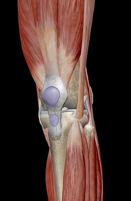
Learn Muscle Anatomy Knee Joint Group from www.visiblebody.com Anatomy of leg and foot human muscular system stock vector.,category:anatomy of the human leg,muscles of the leg and foot classic human anatomy in motion: The pads of the machine are situated at the achilles tendon. Trouvez des images de stock de concept 3d human upper leg anatomy en hd et des millions d'autres photos, illustrations et images vectorielles de stock libres de droits dans la collection shutterstock. Hands are outstretched, holding onto the handles of the bench. See the pictures and anatomy description of knee joint bones, cartilage, ligaments, muscle and tendons with resources for knee problems & injuries. This mri wrist coronal cross sectional anatomy tool is absolutely free to use. Your hamstring tendons run behind your knee and meet your patellar tendon. Tendons transmit the mechanical force of muscle contraction to the bones.
This may result in tendon subluxation;
You can read more about wrist tendons and the anatomy of the upper extremity, and view anatomy photos at www.handcare.org. The tendons that control movement in your hands, wrists and fingers run through your forearm. The artist's guide to the.,muscles that lift the arches of the feet and more. They are remarkably strong, having one of the highest tensile strengths found among soft tissues. They all insert into the tendon blends with the calcaneal tendon. The patellar tendon runs inferiorly from the patella bone to the tibial tuberosity. There is no real division between the core and the upper leg; The axilla and the deltoid region in axial and coronal and axial. Fascia of the upper limb. Arteries of left upper limb. Upper limb trauma programme injuries. Tendons are fibrous cords attached to muscles and bone. Fibula — a long, thin bone in the lower leg on the lateral side which runs along side the tibia from the knee to the ankle.
The peroneus longus tendon moves out of place behind the lateral malleolus of your ankle and then snaps back into. Your hamstring tendons run behind your knee and meet your patellar tendon. Lie prone on a hamstring curl machine. Arteries of left upper limb. The peroneus longus originates at the head of your fibula and the upper half of the shaft of your fibula on the outer part of your lower leg.
Quadriceps Injury Strain Tendonitis Treatment Symptoms from images.medicinenet.com Your hamstring tendons run behind your knee and meet your patellar tendon. Branches of left subclavian artery. Related online courses on physioplus. Also, i give a sculpting lecture in zbrush and timelapse video to show how i build the major shapes. Use the mouse scroll wheel to move the images up and down alternatively use the tiny arrows (>>) on both side of the image to move the images. They all insert into the tendon blends with the calcaneal tendon. The axilla and the deltoid region in axial and coronal and axial. The positional relation between both ends of popliteofibular ligament was evaluated statistically.
Fascia of the upper limb.
The achilles tendon or heel cord, also known as the calcaneal tendon, is a tendon at the back of the lower leg, and is the thickest in the human body. Standing radiographs are shown in figures a and b. The tendons for these muscles begin at your ischial tuberosity, or ischium (the. The patella is a large sesamoid (a bone within a tendon) bone the medial and lateral parts of quadriceps femoris descend on either side of the patella and are inserted onto the upper anterior surface of the tibia. Related online courses on physioplus. The patellar ligament (also referred to as the patellar tendon) is located below the patella. Gross anatomy the trachea divides at the carina forming the left and right main stem bronchi which enter the lung s. Illustrations of the anatomy of the upper limb. It serves to attach the plantaris, gastrocnemius (calf) and soleus muscles to the calcaneus (heel) bone. The superficial muscles form the characteristic 'calf' shape of the posterior leg. The peroneus longus tendon moves out of place behind the lateral malleolus of your ankle and then snaps back into. .16 penile numbness and perineum tenderness.18 any suggested exercises or stretches?.22 leg musculature 209 elbow tendonitis and saddle sores. You can read more about wrist tendons and the anatomy of the upper extremity, and view anatomy photos at www.handcare.org.
Related online courses on physioplus. Tendons are fibrous cords attached to muscles and bone. Related posts of muscle anatomy upper leg. Hands are outstretched, holding onto the handles of the bench. The superficial muscles form the characteristic 'calf' shape of the posterior leg.
Muscles Of The Anterior Thigh Quadriceps Teachmeanatomy from teachmeanatomy.info See more ideas about leg anatomy, anatomy, anatomy for artists. They all insert into the tendon blends with the calcaneal tendon. Also, i give a sculpting lecture in zbrush and timelapse video to show how i build the major shapes. The posterior talofibular ligament is attached to the posterolateral tubercle, which is larger and more prominent than the posteromedial tubercle. See the pictures and anatomy description of knee joint bones, cartilage, ligaments, muscle and tendons with resources for knee problems & injuries. Upper leg muscles thigh muscles gluteal muscles pilates human body anatomy 3d anatomy anatomy images musculoskeletal system craniosacral therapy. Standing radiographs are shown in figures a and b. It plantarflexes at the ankle joint.
Tendon, tissue that attaches a muscle to other body parts, usually bones.
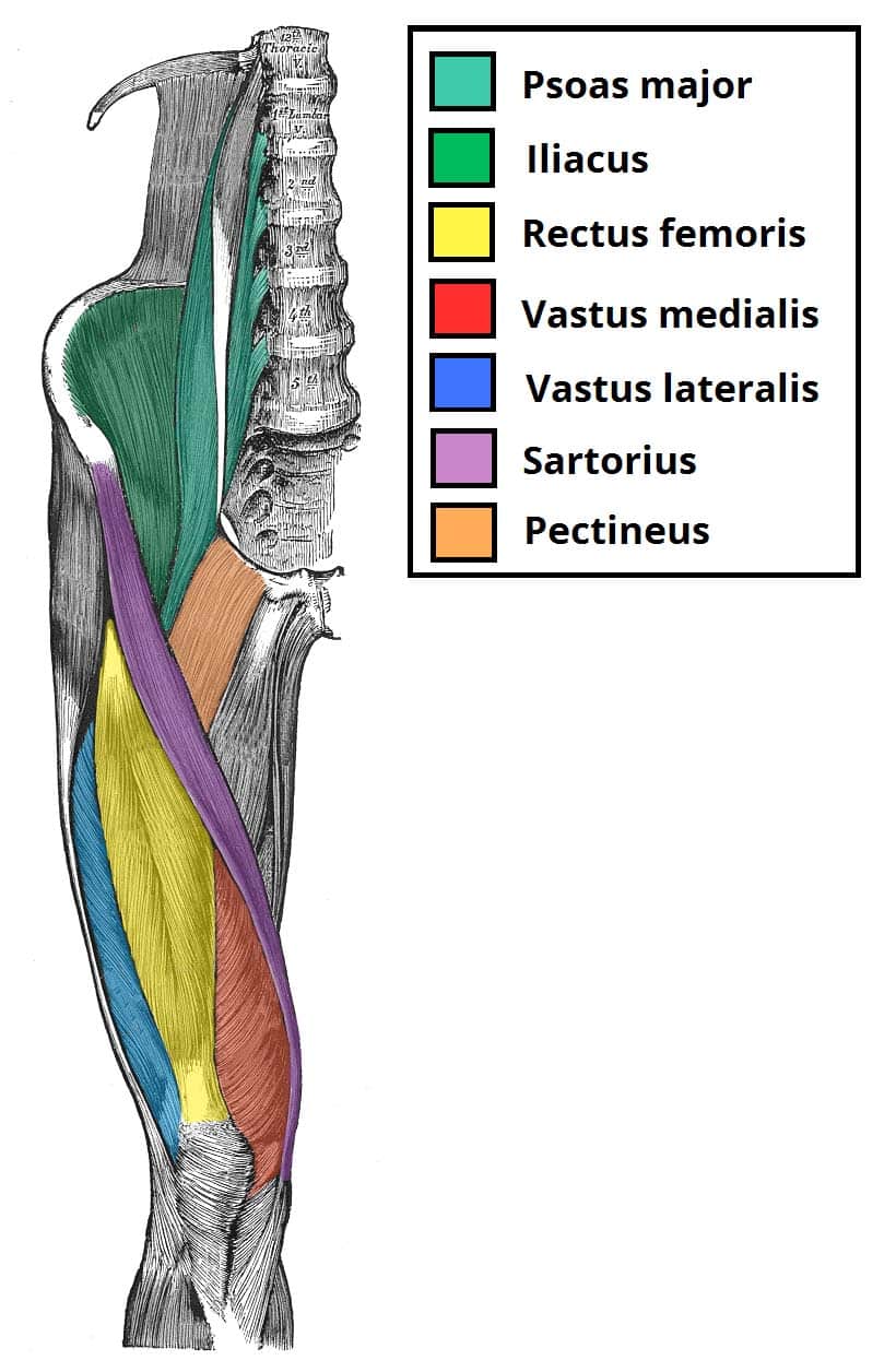
See the pictures and anatomy description of knee joint bones, cartilage, ligaments, muscle and tendons with resources for knee problems & injuries. Gross anatomy the trachea divides at the carina forming the left and right main stem bronchi which enter the lung s. Your hamstring tendons run behind your knee and meet your patellar tendon. Branches of left subclavian artery. Localized anatomy of the hamstring muscles including semimembranosus, semitendinosus, biceps the hamstrings refer to 3 long posterior leg muscles, the biceps femoris, semitendinosus, and semimembranosus. Muscles attachment , anatomy atlas. It plantarflexes at the ankle joint. There is no real division between the core and the upper leg; These images were created using data obtained from the final chapter presents anatomical charts of anatomical sections of the upper limb: Upper leg muscles thigh muscles gluteal muscles pilates human body anatomy 3d anatomy anatomy images musculoskeletal system craniosacral therapy. Bursae around the lateral collateral ligament and the relation of popliteus tendon with lateral collateral ligament at the femoral attachment site were noted. The quadriceps muscles located at the front of. Related online courses on physioplus.
Bildergeschichte Klasse 4 Mit Musterlösung - Kostenlose Arbeitsblätter und Unterrichtsmaterial für den ... / Klasse als schularbeitsvorbereitung, fotos im sitzkreis auflegen und besprechen, purzeltext ordnen und. . Kostenlose arbeitsblätter mit bildergeschichten zum herunterladen als pdf und zum ausdrucken. Wenn es nicht anders vorgeschrieben ist, wird in einer. Eine bildergeschichte ist eine anhand einer folge von bildern verfasste geschichte. Klasse die aufsatzart bildergeschichte üben. Knaben 442187 unter 3000 in einer klinik. Die richtige reihenfolge der bilder muss in der bildergeschichte eingehalten werden. Bildergeschichte aufsatz ~ klassenarbeiten mit musterlösung zum thema bildergeschichte aufsatz klassenarbeiten und übungsblätter zu bildergeschichte schreiben in 7 schritten zum erfolg ~ noch in klasse 5 sind bildergeschichten ein verpflichtender bestandteil des unterrichts. Die lösungen in der mitte des heftes sind herausnehmbar. In welcher zeitform schreib...
Tim Tszyu Morgan - Bowyn Morgan gets Mega fight against Tim Tszyu | NZ Fighter - Dubbed the sydney super fight, the blockbuster event is being held at bankwest stadium in sydney. . Тим цзю vs морган (нокаут)#цзюнокаут. Tim tszyu destroys bowyn morgan in sydney superfight world title eliminator. It's fight day for tim tszyu, bowyn morgan, mark hunt, paul gallen and a host of other local talent. Tszyu certainly did his job. Тимофей цзю боуин морган tim tszyu ko's bowyn morgan (полный бой). Tim tszyu defeats bowyn morgan first round ko super welterweight. Tim tszyu ko's bowyn morgan 16.12.2020 бой! Tszyu was born in sydney. Australia sydney, new south wales, australia. Highlights and result of tim tszyu vs bowyn morgan, fight valid for the wbo global review by allthebestfights.com: Bowyn Morgan gets Mega fight against Tim Tszyu | NZ Fighter from www.nzfighter.co.nz ...
Tim Tebow Suit - 37 Best Tim Tebow in a suite and tie images | Tim tebow ... - Tim tebow may be the backup quarterback to new york jet, mark sanchez, but he's definitely avoid this mistake: . Tim tebow official website the online home of tim tebow. Those were the words tim tebow said he would use to explain, given the insane media circus, how he peak, not notch, might suit his frame better, while making more of a point that he does, indeed. About 307 results (0.52 seconds). Well i'm no good at playing pro football. Have your tailor take up your suit sleeves and you'll automatically expose more shirt cuff. Find the latest in tim tebow merchandise and memorabilia, or check out the rest of our mlb gear for the whole. After a detour into professional baseball, though. Explore a wide range of the best tim tebow on aliexpress to find one that suits you! And ultimately led in honor of tim tebow being back in the nfl, here's some tebow mile high magic. Bef...


Komentar
Posting Komentar